The Body in Conversation: Part 5-Rooted in the Spine
When we think about the diaphragm, most of us picture a dome sitting underneath the lungs.
Maybe we imagine it dropping down as we inhale and rising back up as we exhale.
This is a useful basic image, but remarkably incomplete. So, let's flesh it out.
The diaphragm doesn’t just hang beneath your ribs like a soft parachute. or like a coffee plunger which is how I most often see it depicted.
When we follow its route backwards and downwards things get much more interesting.
The diaphragm is firmly attached to your ribs, your sternum and, very crucially, your spine.
It reaches down into the lumbar region of the spine through strong bands of muscle known as the crura, and it forms relationships with muscles and tissues that most people would never think of as having anything to do with breathing.
Your psoas and your quadratus lumborum, which we will chat about in more detail a little later. The fascia around the lower back and the structures travelling between your chest and abdomen.
As you can start to surmise the diaphragm is not simply sitting at the top of the abdominal cavity, pumping up and down.
It is actually woven into the very architecture of your trunk.
And every time you breathe, that bio-mechanical architecture moves.
The crura: where the diaphragm meets the spine
At the back of the body, the diaphragm narrows into two muscular and tendinous pillars called the right and left crura.
Crus simply means “leg”.
The right crus generally extends further down the lumbar spine than the left, attaching all the way down to the upper lumbar vertebrae and their discs.
This immediately changes the picture of the diaphragm.
It isn’t just a dome, It has roots and those roots connect breathing directly into the spinal region.
As you breathe, the diaphragm changes shape and tension. Its central tendon moves, its muscular fibres shorten and lengthen, the lower ribs move and the pressure relationships between the chest and abdomen change.
And all of this happens while part of the muscle is anchored into the lumbar spine.
That doesn’t mean the diaphragm is yanking your vertebrae around with every breath.
The body is much more subtle than that.
But it does mean breathing and spinal mechanics cannot really be separated into two completely different systems.
They are already physically connected.
Your diaphragm bridges over other muscles
This is one of my favourite bits of diaphragm anatomy.
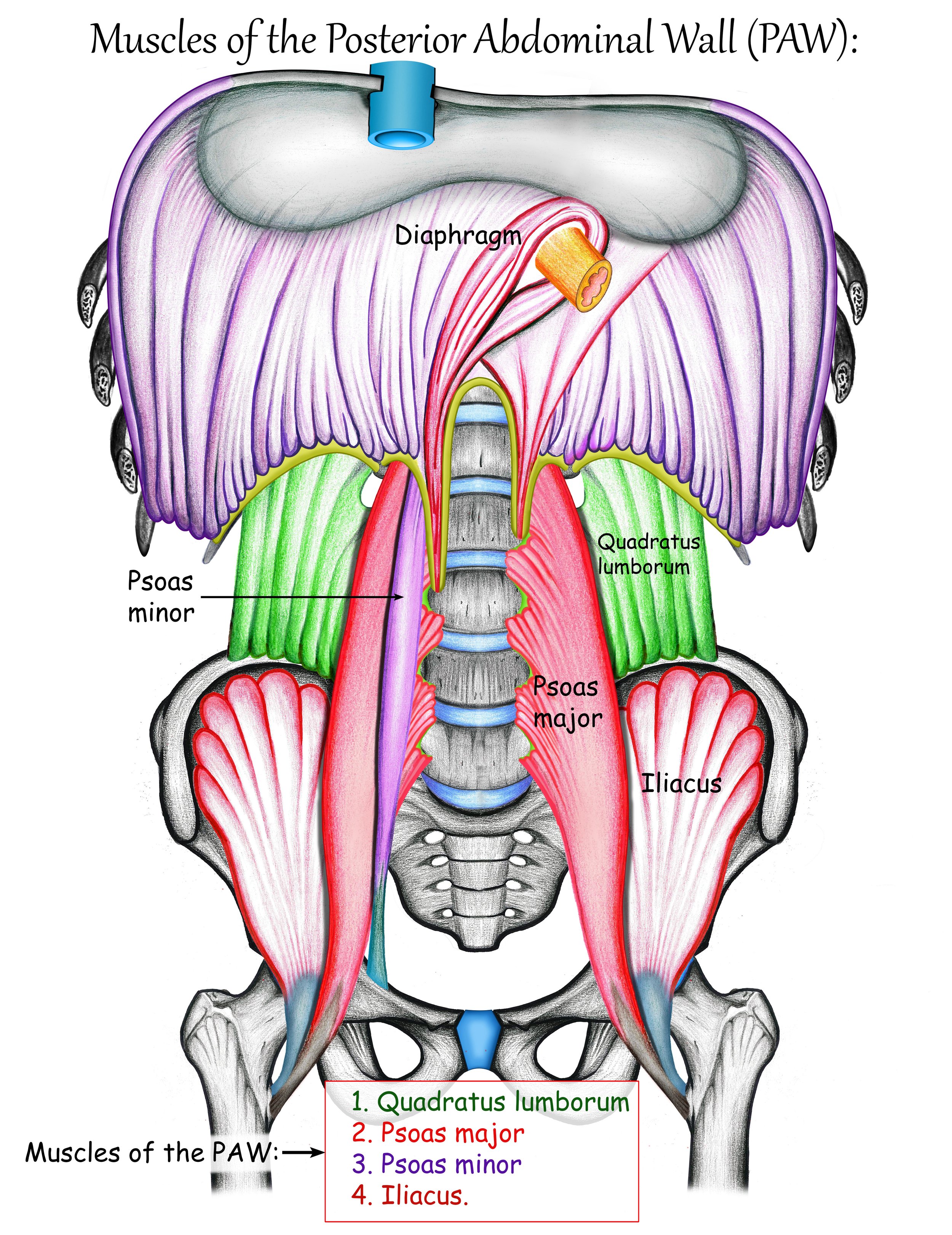
As the diaphragm travels around the back of the body it forms arch-shaped connective tissue structures called the arcuate ligaments.
One of these arches passes over the psoas major and another passes over quadratus lumborum.
So instead of visualising the diaphragm, psoas and QL as separate muscles neatly stacked beside each other, it may be more useful to imagine a neighbourhood.
They have their own jobs.
They have their own attachments.
But they share walls, borders and connective tissue.
The diaphragm literally spans across them.
Now you can start to really see that the way you breathe can and does change depending on how you organise your spine, hips and trunk.
The psoas connection
Psoas has achieved almost mythical status on the internet.
It has been called lots of different names;
The muscle of the soul.
The trauma muscle.
The fight-or-flight muscle.
The emotion muscle.
The hidden reason for everything from back pain to anxiety.
There is some fascinating anatomy here.
But we don’t need mythology to make it interesting.
Psoas major originates from the lumbar spine and travels down through the pelvis to attach to the femur.
It participates in hip movement and influences the relationship between the spine, pelvis and legs.
And near the top of that journey, the diaphragm arches over it.
They also sit within a region full of fascia, blood vessels, nerves and other structures.
But anatomical proximity does not automatically mean that one muscle “controls” the other.
And it definitely does not mean that releasing your psoas will magically release stored trauma, fix your diaphragm or reset your nervous system.
The real relationship is both less dramatic and way more interesting.
If you change the organisation of your pelvis and lumbar spine then the mechanical environment around the diaphragm changes.
If you change the way you breathe then the pressure, shape and movement occurring through the trunk will change too.
Neither structure works in isolation.
Quadratus lumborum: the quiet neighbour
Quadratus lumborum, or QL, sits at the back of the abdominal wall.
It connects the pelvis, lumbar spine and lowest rib.
Its relationship with the diaphragm is particularly interesting because the diaphragm's lateral (which means side) arcuate ligament passes over it.
The QL is often described simply as a side-bending muscle, but like most muscles its function changes depending on position and which parts of the body are fixed.
It contributes to stability of the lumbar region and can influence the position and movement of the twelfth rib, which you can think of as being roughly around the lower edge of the rib cage rather than using it as an exact surface landmark.
And that twelfth rib is also part of the diaphragm's attachment.
If the ribs move differently, the diaphragm is working from a different shape.
If the spine is rigid, rotated or held in one strategy all day, the diaphragm still has to breathe within that strategy.
If the body is constantly bracing through the back of the trunk, breathing doesn't stop.
It just has to adapt.
Breathing is a three-dimensional event
This is why I keep coming back to the idea of the ribs moving like gills.
Breathing is not simply the belly pushing forwards.
Nor is it just the chest lifting.
Nor is it just the diaphragm pumping up and down like a coffee plunger.
As we inhale, the rib cage changes shape in multiple directions.
The lower ribs widen.
The back of the rib cage moves.
The abdomen lengthens and expands.
The diaphragm changes shape.
The pelvic floor responds to the changing pressure above it.
And the spine participates within that movement.
If you lie on your back and think only about pushing your tummy upwards, you are seeing just one small part of the system.
Try imagining the back of your lower ribs as well.
Can they widen?
Can the breath move behind you?
Can you sense the area where your ribs begin to become your back?
For some people this feels surprisingly difficult.
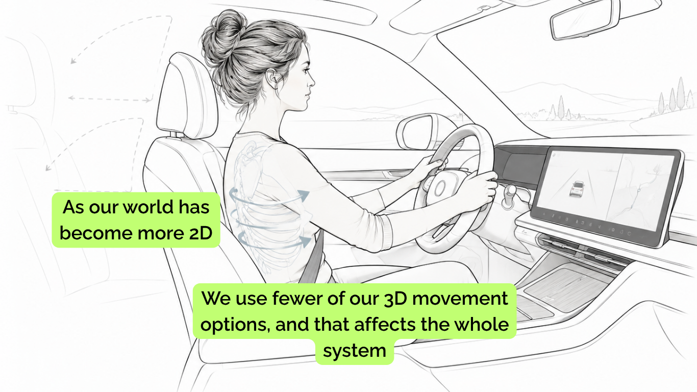
Not because something is wrong, but in part because our view of the world has become more and more 2D. In some high-end cars, like my brother-in-law's, you don't even have to look in the side mirrors because the whole navigation system is in front of you. There is less and less need to rotate your neck or thoracic cage and actually look around you.
Our ergonomic dream of having everything conveniently placed directly in front of us may also mean that, for many of us, the back and sides of the body have simply become areas we don't pay much attention to anymore.
The diaphragm and posture
This is where conversations about posture can quickly become quite unhelpful.
There is actually no single perfect position in which your diaphragm suddenly works properly.
Bodies are meant to move.
We are designed to bend.
To Rotate.
To Slouch.
To Reach.
To Run.
To Curl up on the sofa with a good book or a blog :)
To Carry children.
To Lift weights.
And we often sit at desks.
The question is not whether you maintain the perfect position all day.
The more useful question is:
Do you have options?
Can your ribs expand in different directions?
Can your spine flex and extend?
Can your pelvis move underneath your rib cage?
Can you rotate?
Can you breathe while you are doing those things?
Or has your body found one strategy and decided to stay there?
Because if the trunk becomes very rigid, the diaphragm doesn't stop working.
It simply has fewer movement options available around it.
And other parts of the respiratory system may have to contribute differently.
Why your back moves when you breathe
Place your hands around your lower ribs.
Thumbs pointing towards your spine.
Just take an easy breath in.
Don't take the biggest breath you can.
Take a moment to notice.
Can you feel movement underneath your thumbs?
Now visualise the back of the diaphragm widening inside you.
Not forcing.
Not pushing.
Just making space.
This posterior movement is one of the reasons I dislike the instruction to simply “breathe into your belly”.
The breath is not going into your belly.
Air remains inside your lungs.
What you are feeling is the mechanical response of the body to the diaphragm contracting, the lungs filling and the rib cage and abdominal wall changing shape.
And some of that movement should be available behind you.
Your back is part of your breath.
The fascial conversation
This is also where fascia becomes difficult to ignore.
The diaphragm exists within a continuous network of connective tissue.
Its fascia blends and communicates with neighbouring tissues.
The structures around the spine, psoas, QL, abdominal wall and thorax are not wrapped individually in vacuum-sealed plastic bags.
Dissection and anatomy textbooks do understandably separate structures because we need to learn their names.
Real bodies are much less tidy.
They slide.
They pull.
They transmit force.
They share connective tissue.
This is why models such as Thomas Myers' Deep Front Line from his book and teaching called Anatomy Trains can be super useful.
The Deep Front Line describes a continuous pathway running through structures including the deep foot, inner leg, pelvic floor, psoas region and diaphragm.
It can be a brilliant way of thinking about movement continuity.
But it is important to remember what it is.
A model.
It is not an anatomical railway line running through the body with force travelling neatly from one station to another.
The model helps us ask questions about how those structures might cooperate and work together.
Connection does not mean causation
This matters because fascia has become one of those subjects where anatomical relationships are sometimes stretched much further than the evidence allows.
Yes, the diaphragm is anatomically connected to structures around the spine.
Yes, fascia provides continuity throughout the body.
Yes, breathing changes pressure and movement through the trunk.
But that doesn't mean every restriction in the hip originates in the diaphragm.
It doesn't mean a tight psoas is preventing you from breathing.
And it doesn't mean one clever release technique can reset the entire system (sadly- that would be so cool if it did).
There absolutely is a connection.
Here is what that connection actually is.
Here is what we can reasonably infer from it.
And then there is the point where people have run away with it.
I am interested in the first three.
Try this: breathing into the back of the body
Read this and then take a moment to do the exercises below:
Lie on your back with your knees bent, or sit somewhere comfortable.
Place your hands around your lower ribs so your fingers are at the sides and your thumbs reach towards the back.
Take a few of your regular breaths.
Notice what happens underneath your hands.
Don't deliberately make your breath enormous.
Instead imagine the lower ribs widening sideways and slightly backwards, almost like a pair of gills opening.
Now notice your lower back.
Does it feel rigid?
Does it subtly change shape as you breathe?
Can you allow the back of your body to receive the breath rather than trying to push the breath forwards?
Stay there for four or five breaths.
Then stand up.
Take another breath, has anything changed.
Perhaps nothing major has but also conversely perhaps it has. Or may there has been a little shift in your awareness.
There is no right or wrong here, no "you should have felt X, Y, Z".
We are not trying to fix anything.
We are just learning to notice.
Try this: let the spine join the breath
Stand comfortably with your feet underneath you.
Take a breath in and gently allow your chest and spine to lengthen, the curve in your lower back to deepen a little, notice if you can your tailbone wanting to stretch out behind you.
As you breathe out, let yourself soften back to where it was.
Nothing too exaggerated just notice the movement pattern.
The point is to experience that breathing and spinal movement are capable of adapting to one another.
Your diaphragm isn't waiting for you to find the perfect posture.
It is moving with you.
Your diaphragm has roots
Perhaps that is the biggest shift in how I would like us to think about this muscle.
The diaphragm is not an isolated breathing dome floating in the middle of the body.
It has ribs around it.
A heart above it.
A liver underneath it.
Organs moving beneath it.
Blood vessels and an oesophagus passing through it.
A spine behind it.
Muscles beside it.
Fascia continuing away from it.
Every breath reaches further into the body than you think.
Your diaphragm doesn't just move air.
It participates in movement.
And its roots run all the way into your spine.
But of course, if this huge moving structure forms the roof of your abdominal cavity, there is another question we need to ask.
What happens underneath it when it moves?
Because every time the diaphragm descends, changes shape and reorganises the space inside your trunk, pressure has to go somewhere.
And below it sits another extraordinary moving structure that is very often taught as though it were working entirely on its own.
Your pelvic floor.
That is where we are going next.