The Three Gateways
Through, Within and Behind the Diaphragm
So far, we have looked at the diaphragm as a constantly in motion landscape.
Two slightly different jellyfish domes changing shape beneath the lungs.
A central tendon intimately related to the pericardium surrounding the heart.
Ribs opening and recoiling like gills around its muscular edges.
But there is another huge problem this extraordinary structure has to solve. The diaphragm forms a substantial boundary between the chest and the abdomen. And yet some very important structures need to travel from one side of that boundary to the other.
The blood needs to return to the heart.
It is pretty essential too for food to reach the stomach.
The enormous aorta needs to carry blood down from the heart into the abdomen and beyond.
So the diaphragm needs gateways, portals, from one realm to another.
And turning yet again to the engineering of our body, it has not simply punched three identical holes through itself.
Each one of these gateways is constructed completely differently.
One passes through the central tendon.
One passes through muscle.
And one actually travels behind the diaphragm.
Same border guard.
Three very different solutions.
Imagine three travellers approaching a border
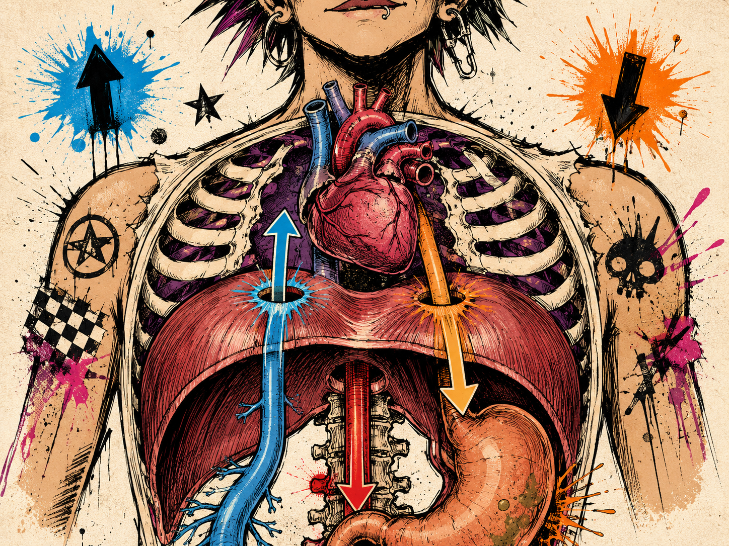
Picture the diaphragm as a moving border between two busy countries.
Above it: the thorax.
Below it: the abdomen.
Now imagine three travellers arriving.
The first is the inferior vena cava, carrying blood back towards the heart. It is given a carefully reinforced opening through the central tendon of the diaphragm.
The second is the oesophagus, carrying everything you swallow towards the stomach. Its route passes through a muscular gateway formed largely by fibres associated with the right crus of the diaphragm.
The third is the aorta, the enormous artery carrying blood away from the heart. And its solution is different again. Rather than travelling through the central tendon or directly through the contracting muscular belly of the diaphragm, the aorta passes posteriorly (to the back) between the two crura (the two arms of the diaphragm that anchor into the spine) beneath the median arcuate ligament.
Already, I think that's rather brilliant.
Three structures crossing the same anatomical boundary in three completely different ways.
Gateway One: The Inferior Vena Cava
Let's start with the inferior vena cava, or IVC.
This is one of the largest veins in your body. Its job is to collect deoxygenated blood from the lower half of the body and return it upwards towards the heart.
And where is the heart?
Of course we already know that it is immediately above the diaphragm.
The IVC travels upwards from the abdomen, passes through an opening in the diaphragm and enters the right atrium.
Its opening, the caval opening, lies within the diaphragm's central tendon, classically around the level of T8 (one of your thorax vertebrae).
Notice what is unusual about this.
It passes through tendon rather than contracting muscle.
The vessel is therefore intimately integrated with a part of the diaphragm that changes position as the surrounding muscular fibres contract and relax.
At the same time, breathing alters pressure within the chest and abdomen.
So breathing and circulation are not occurring in completely separate mechanical worlds. They work in tandem.
Another important biological conversation.
A window in the central tendon
To make this easier to visualise, I picture this first gateway almost like an eyelet sewn into strong fabric.
The central tendon is the fabric.
The vena cava passes through the eyelet.
As the diaphragm changes shape, the landscape surrounding that opening moves with it.
Not a random hole punched through tissue.
A vessel travelling through a moving structure.
And because the IVC passes through the diaphragm immediately before entering the heart, breathing and circulation meet here in a very practical way.
As you inhale, pressure inside the chest falls while pressure in the abdomen changes. That pressure difference can help encourage venous blood to travel upwards towards the heart, part of what is sometimes called the respiratory pump.
So every breath is doing more than moving air.
It is also subtly changing the pressure in the spaces through which your blood is returning to your heart.
Gateway Two: The Oesophagus
The second traveller behaves very differently.
Your oesophagus has a really important job to do too. It carries food and fluid from your throat down towards your stomach.
To get there, it must pass from the thorax into the abdomen.
But it doesn't pass through the central tendon.
It travels through a muscular opening called the oesophageal hiatus, classically around T10. The opening is intimately related to the diaphragmatic crura, particularly fibres of the right crus.
The anterior and posterior vagal (vagus nerve) trunks also pass through this region.
So now our gateway isn't a tendinous eyelet, no not this time. this time it is surrounded by living, contracting muscle.
And this difference is necessary.
Imagine a soft drawstring doorway
This is how I like to picture the oesophageal hiatus.
Imagine the oesophagus travelling through a soft muscular drawstring doorway.
Not tightly strangling it, never clamped shut, always super responsive. Capable of subtly changing shape and tension as the diaphragm contracts and relaxes.
Anatomical studies of this region show that the hiatus (gap) itself changes shape with breathing, with contraction of the diaphragmatic crura helping the narrowing around the oesophagus during deeper inspiration.
This muscular contribution forms part of the wider mechanism that helps prevent stomach contents from travelling back upwards into the oesophagus.
So the gastro-oesophageal junction (the exact meeting point where your food pipe connects to your stomach) is not simply: a tube with a valve. The diaphragm has a part to play too..
The fascia around the gateway
And there is another important structure here. The oesophagus is not simply hanging freely inside a muscular hole.
Around it sits the phrenoesophageal ligament, sometimes called the phrenoesophageal membrane. This connective-tissue structure helps attach the oesophagus to the diaphragm around the hiatus.
Anatomical studies describe it as arising from fascial layers above and below the diaphragm - including the endothoracic and transversalis fascia - and inserting around the oesophagus. It contains collagen and elastic fibres, allowing it to anchor the region while still accommodating movement during breathing and swallowing.
To actually picture this, imagine the oesophagus as a soft flexible tube dropping down through the centre of a stretchy, muscular doorway.
Now add a second layer.
Wrapped around the oesophagus is the phrenoesophageal membrane - a thin, elastic layer of connective tissue. I like to imagine it almost like a soft collar or little flared skirt, gently attaching the oesophagus to the diaphragm around the edges of the opening.
Outside that sits the muscle of the diaphragm itself.
So the full picture is this:
the oesophagus travelling down through the middle;
a soft elastic collar of fascia embracing it;
and the muscular fibres of the diaphragm forming the moving doorway around them both.
As you breathe, none of these structures stays completely still. The diaphragm changes shape. The muscular opening subtly changes with it. The oesophagus needs enough freedom to glide when you swallow. And that elastic fascial collar helps keep everything connected while still allowing that movement to happen.
It is less like a pipe pushed through a rigid hole...
and much more like a flexible tube passing through a soft, responsive, elastic sleeve - all moving together.
Once again:
there are no neat edges.
When the Gateway Changes: Hiatal Hernia
This is where the anatomy becomes particularly relevant to something I see regularly in practice:
hiatal hernia.
In a sliding hiatal hernia, the gastro-oesophageal junction and part of the upper stomach move upwards through the oesophageal hiatus into the chest.
The anatomy of this region can change in several ways.
The hiatus (gap) may widen.
The relationship between the oesophagus and crural diaphragm may change.
The phrenoesophageal connective tissues may become stretched or altered.
And the lower oesophageal sphincter and crural diaphragm may no longer sit in quite the same relationship to one another. Hiatal hernia is multifactorial, so it would be far too simplistic to blame one structure alone.
But understanding the local anatomy helps us understand why reflux can so often accompany it.
Two parts of the anti-reflux system
One of the things I find fascinating about this region is that there are really two overlapping contributors to the pressure barrier at the gastro-oesophageal junction.
There is the lower oesophageal sphincter, which belongs to the oesophagus itself.
And there is the crural diaphragm, whose muscular fibres surround the oesophageal hiatus and can contribute extra pressure from outside.
So your diaphragm is not simply sitting next to the digestive tract.
Part of it is actively participating in this gateway.
Which brings us to a question I am asked a lot.
Can breath work help a hiatal hernia or reflux?
This is where the answer needs a little nuance.
Breathing exercises cannot make a promise or claim to pull a structural hernia back into place, shrink the hiatus or magically tighten stretched fascia. Changes in fascia take months and even years.
But that doesn't mean breathing mechanics are irrelevant actually it is quite the opposite.
The crural diaphragm is muscle and Its function contributes to the gastro-oesophageal pressure barrier.
Breathing changes pressure through the thorax and abdomen.
And the oesophageal hiatus (the gap) itself responds dynamically to diaphragm contraction.
Research into diaphragmatic breathing in people with GERD has shown improvements in lower oesophageal sphincter pressure and reductions in reflux events in some groups. A 2020 randomised study, for example, found that diaphragmatic breathing increased inspiratory lower oesophageal sphincter pressure and reduced post-meal reflux events.
What I have witnessed with my clients over the years
This is also where my work an lived experience becomes relevant.
I see marked improvements in reflux symptoms in many of my clients with GERD and hiatal hernias when we work on breathing mechanics.
Often we are not simply teaching someone to “take deeper breaths”.
We are looking at the whole strategy.
Can the lower ribs move more freely?
Can the diaphragm change shape without the person forcing the abdomen forwards?
Are they constantly bracing?
Do they hold their breath during movement?
Are they creating large spikes in abdominal pressure unnecessarily?
Can the rib cage open and recoil more easily?
Can we improve coordination around the diaphragm rather than continually asking the abdominal wall to grip?
And very often, symptoms change.
That does not necessarily mean the anatomical hernia has disappeared.
Structural change and functional improvement are not the same thing.
A person can experience a very meaningful reduction in symptoms because the way the system is functioning has changed, even if the underlying structural anatomy remains.
That distinction is really important.
And it means the more useful question is probably not:
“Can I breathe my hernia away?”
but:
“Can I improve how this whole region moves, coordinates and manages pressure?”
That is something we can explore.
Bring back the soft collar
Come back to our image.
Imagine the oesophagus travelling down towards the stomach.
Around it is that soft elastic connective-tissue collar - the phrenoesophageal membrane.
Surrounding the gateway are the muscular fibres of the crural diaphragm.
Now imagine breathing without shoving anything.
The lower ribs open gently like the gills we explored in Chapter Three.
The two diaphragm domes change shape beneath the lungs.
The muscular gateway around the oesophagus responds as part of that movement.
The abdominal contents adapt below.
Then the breath releases and everything recoils.
Nothing needs to be yanked down.
Nothing needs to be “put back”.
We are encouraging movement, coordination and pressure management around a region designed to move.
That is a much more useful image.
Your breathing muscle is also in swallowing territory
Think about how extraordinary this is.
We tend to categorise:
breathing
and
digestion
Like they belong to completely separate departments.
But here is your oesophagus travelling through fibres belonging to the primary muscle of respiration.
And travelling alongside it are the vagal trunks - major neural pathways between the brain and viscera (organs).
That does not mean every digestive problem is caused by the diaphragm.
It also does not mean one breathing exercise “resets your vagus nerve”.
And it does not mean reflux is always a breathing problem.
But it does mean that breathing, swallowing, digestion, neural communication and pressure occupy shared anatomical territory.
That is worth knowing.
Gateway Three: The Aorta
And then we get to my favourite one.
The aorta.
The enormous artery leaving the heart and carrying oxygenated blood down through the body.
It descends through the thorax towards the abdomen.
You might reasonably assume that it needs another hole through the diaphragm and you would be right.
But its route is different again.
The aortic hiatus sits posteriorly (towards the back) between the two diaphragmatic crura, close to the vertebral column and beneath the median arcuate ligament, classically around T12.
The thoracic duct and azygos vein also travel through this region.
So rather than travelling through the central tendon or through a muscular opening like the oesophagus, the aorta takes a protected posterior route.
Imagine a tunnel beneath a moving bridge
For this one, imagine a road tunnel running beneath a moving bridge.
The diaphragm can change shape above it.
The crura can move.
Breathing continues.
But the aorta still needs to deliver blood uninterrupted.
Anatomical studies of the aortic hiatus suggest that this region is arranged so that diaphragmatic contraction does not simply act like a tight muscular ring compressing the aorta.
Again, the architecture fulfills the job description perfectly.
Through. Within. Behind.
This is the bit to remember.
The vena cava:
through the central tendon
The oesophagus:
through a muscular gateway
The aorta:
behind the diaphragm, between the crura
Three structures.
Three routes.
And when you see them together, the diaphragm becomes much harder to imagine as a simple sheet of muscle pumping mechanically up and down.
It is architecture, gorgeous Moving architecture.
And there are more than three holes
Before somebody anatomically minded emails me - yes, there are other structures crossing the diaphragm too.
Nerves, vessels and lymphatic structures all need routes between the chest and abdomen.
The sympathetic trunks, splanchnic nerves and smaller vessels have their own pathways.
So when I say The Three Gateways, I mean the three major openings traditionally used to teach diaphragmatic anatomy.
The reality, as always, is much more intricate.
Because bodies stubbornly refuse to organise themselves according to the chapter headings in anatomy textbooks.
Remember T8, T10, T12?
If you have ever had to learn diaphragm anatomy, you may have memorised:
IVC — T8
Oesophagus — T10
Aorta — T12
Useful for an exam.
But unless you're studying anatomy, I'm much more interested in whether you remember:
Tendon.
Muscle.
Behind.
Because those relationships tell us much more about what is actually happening.
And now we need to talk about the legs of the diaphragm
Did you notice the word crura keeps appearing? The oesophageal opening is intimately related to them. The aorta passes between them.
But what actually are they?
Crus literally means leg.
The diaphragm has strong muscular and tendinous extensions that descend posteriorly and anchor it towards the lumbar spine.
So those two jellyfish-like domes we imagined in Chapter Two?
They have surprisingly deep roots. They don't simply finish at the bottom of your ribs. They travel backwards and downwards towards your spine.
And that is going to lead us somewhere very interesting.
But I am getting ahead of myself.
That belongs to Chapter Five.
Did You Know?
Your Oesophagus Shares Its Gateway With the Vagus Nerve
Your oesophagus does not travel through its muscular doorway alone.
The anterior and posterior vagal trunks also pass through the oesophageal hiatus on their journey into the abdomen.
The vagus nerve forms a major part of parasympathetic communication between the brain and many of the organs of the chest and abdomen.
So travelling through one tiny anatomical neighbourhood we have:
the tube carrying your food
and
a major neural communication pathway between brain and viscera.
Again, this is not evidence that one breathing trick can “reset the vagus nerve”.
It is simply another beautiful example of how closely our supposedly separate systems share space.
Explore It: Three Pathways Through a Moving Landscape
You cannot feel these three openings directly from outside your body.
And I don't want you poking around trying, it would probably just get sore.
Instead, let’s build an internal picture.
Sit or stand comfortably.
Place your hands around your lower rib cage and remember those soft gills opening around the two diaphragm domes.
Now picture three routes through this landscape.
Imagine the vena cava travelling upwards, through the central tendon and towards your heart.
Imagine the oesophagus travelling downwards, through its muscular doorway towards your stomach.
Then imagine the aorta descending deeply behind the diaphragm, close to your spine.
Take several normal breaths.
Don't try to create a sensation.
Just imagine:
one travelling up.
two travelling down.
three different routes through one constantly shifting terrain.
The goal is not to feel the vessels.
It is to turn a flat anatomy diagram into something three-dimensional.
Explore It: Breath, Swallow, Breath
Take two or three relaxed breaths.
Notice their rhythm.
Now swallow once.
Notice the tiny interruption and reorganisation.
Then allow your breathing to resume naturally.
Try it again.
Breathe.
Swallow.
Breathe.
You do not need to analyse every sensation.
Just notice that swallowing and breathing have to be coordinated.
The oesophagus passes through the diaphragm.
The airway sits nearby.
Pressure is changing.
Muscles are contracting and releasing.
And your body quietly organises the timing without you consciously managing any of it.
If You Have Reflux or a Hiatal Hernia
Try a gentler exploration.
Sit comfortably rather than folding or slumping tightly through your upper abdomen.
Place your hands around the lower ribs.
Take an ordinary inhale and imagine the ribs gently opening like gills.
Think width and space, rather than pushing the abdomen hard forwards.
Allow the breath to release naturally.
Try five relaxed breaths.
The aim is not to drag the stomach down or force the hiatus closed.
It is simply to explore whether your lower ribs and diaphragm can move with less gripping and more ease.
For somebody who habitually braces or breath-holds, that alone can feel totally different.
The diaphragm is becoming less and less like “a breathing muscle”
This is what I hope is beginning to happen as we move through this series.
Chapter One introduced the diaphragm.
Chapter Two turned it into two moving domes with an intimate relationship to the heart.
Chapter Three wove it into ribs that open and recoil like gills.
And now Chapter Four has brought us: a major vein; your oesophagus; the vagal trunks; your largest artery; the thoracic duct; connective tissue surrounding the gastro-oesophageal junction; and a muscular gateway involved in reflux control.
“the muscle you use to breathe”
Is maybe starting to feel like a spectacularly inadequate way to describe it.
The diaphragm sits at a crossroads of: breathing; circulation; digestion; lymphatic flow; neural communication; pressure; and movement.
It doesn’t control all of those systems. But it is sitting right in the middle of their traffic. It has a friendship based non controlling relationship with these super important parts of your body.
Next Week: Rooted in the Spine
Next, we are going backwards.
Because tucked behind everything we've explored so far are the crura - the legs of the diaphragm.
And they travel much further down towards the lumbar spine than many people realise.
We'll explore the lumbar vertebrae, psoas, quadratus lumborum and arcuate ligaments.
This is also where we'll properly bring fascia and Thomas Myers' Deep Front Line model into the conversation - while separating the anatomy we can actually observe from the models we use to help understand it.
If Chapter Two gave our diaphragm jellyfish...
and Chapter Three gave our ribs gills...
Chapter Five is going to give the diaphragm roots.
Welcome further down the rabbit hole 🐇