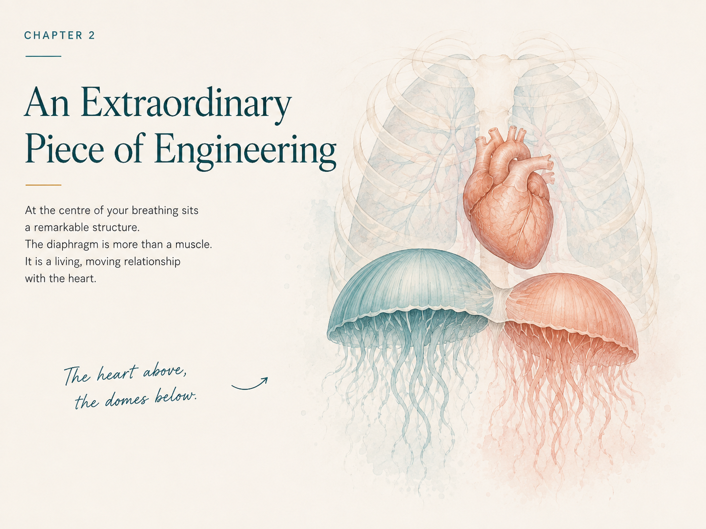
An Extraordinary Piece of Engineering
Diaphragm series - Part 2
The Diaphragm’s Domes, Central Tendon and Its Relationship With the Heart
In Chapter One, we had already started to move away from the idea of the diaphragm as simply the muscle we use to breathe.
Now I want to zoom in a little closer.
Because before we even follow the diaphragm into the ribs, spine, abdomen, pelvic floor, nervous system or fascia, its actual structure is something we need to pause on as it is quite extraordinary.
The diaphragm is not as many folk think a flat sheet stretched across the body.
It is actually a large, three-dimensional musculotendinous structure, shaped into two domes, with muscular fibres travelling inwards towards a strong flat central tendon.
And sitting immediately above that central tendon is the heart.
Well... almost.
Because this is where the anatomy becomes way more interesting.
First, stop picturing a flat diaphragm
A lot of anatomy diagrams do the diaphragm a little bit of a disservice.
It can appear as a curved line separating the chest from the abdomen. This is definately useful for orientation.
Not so useful if you actually want to understand how it moves. In the body, the diaphragm is beautifully three-dimensional.
If you picture in your mind two domes rising upwards into the rib cage.
Around the outside of the domes are muscular fibres.Those fibres travel towards a flatter tendinous area near the centre this is the central tendon.
Above the diaphragm sit the lungs and heart.
Below it sit the abdominal organs.
So the diaphragm forms both:
the floor of the thoracic cavity
and
the roof of the abdominal cavity.
It is living between two enormous moving spaces.
It is not just dropping down or popping back up it is connected and moving as a part of and with the surrounding structures. Which means it will also influence and be influenced by them.
Two domes, not one
We often refer to the diaphragm as though it is perfectly symmetrical.
It isn’t.
It has a right and left hemidiaphragm, and each side occupies a different shape and space
On the right, the liver dominates the space directly beneath the diaphragm, with the gallbladder tucked against its undersurface. Deeper in the upper abdomen sits the pancreas. On the left, the stomach and spleen help create a very different anatomical landscape.
Above, the lungs themselves are also different shapes, and the heart occupies considerable space centrally and towards the left.
So right from the beginning, the diaphragm exists in asymmetry and, that is worth remembering when we look at breathing.
Different does not automatically mean dysfunctional.
Bodies do not have to move like mirrored diagrams. We are not asymmetrically the same.
The two sides of the diaphragm may not sit at exactly the same height or move through exactly the same range.
And their movement will be influenced by what sits above, below and around them.
Even the familiar explanation often given that the right dome sits higher simply because of the liver is probably a little too neat.
As usual, the body is more interesting than the shorthand often afforded to it.
One of the things I like about the Franklin Method is the emphasis on dynamic imagery.
Rather than memorising a fixed picture of anatomy, we can imagine and visualise the structure doing what it actually does.
So instead of picturing the diaphragm as a single dome simply moving up and down, try imagining two slightly different jellyfish floating side by side conjoined inside the lower rib cage.
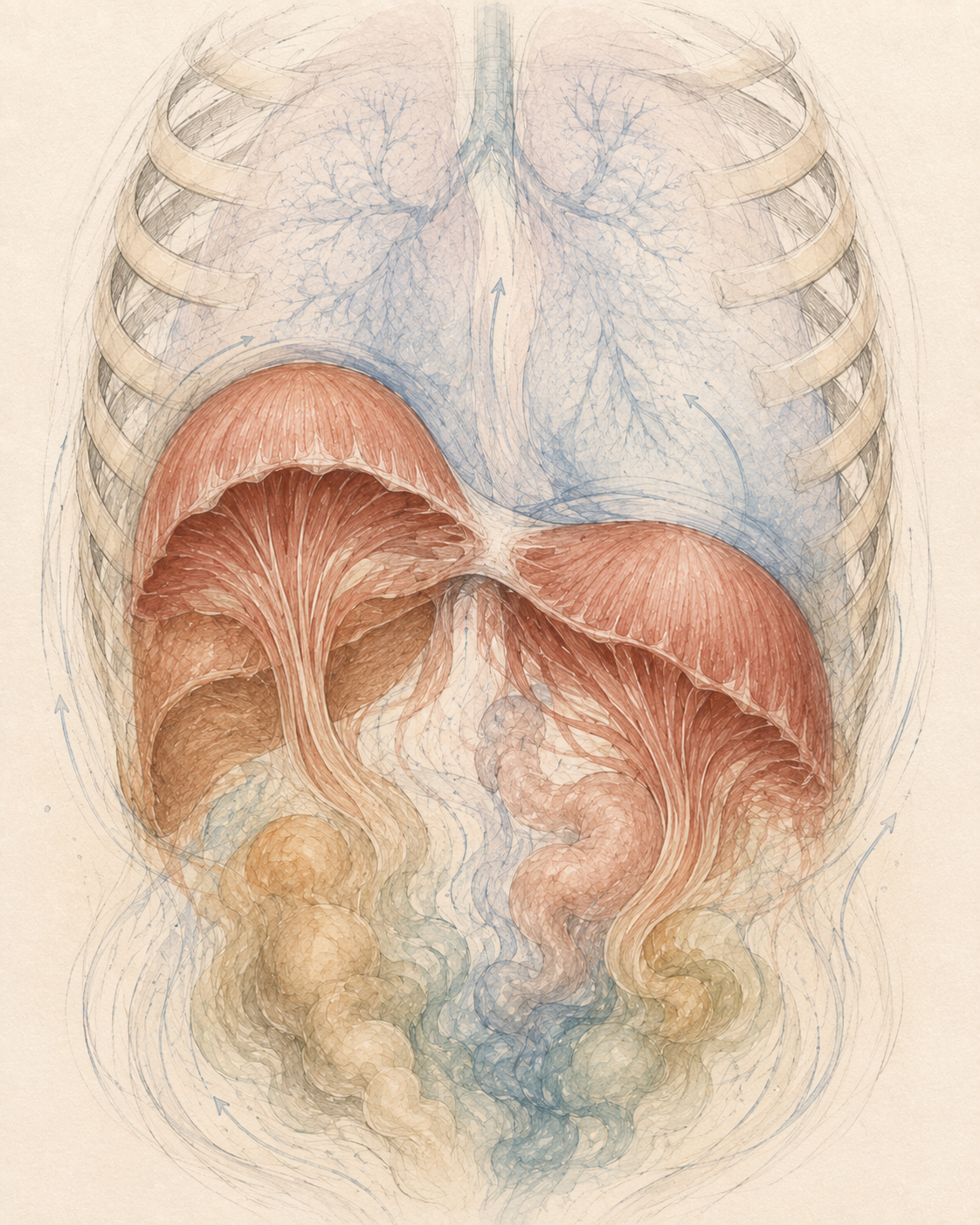
The image to help you
I can really get this when I close my eyes and breathe
Not identical twins.
Two related shapes, each a slightly different shape and size.
As you breathe in, imagine the muscular fibres of each dome shortening and changing shape, a little like the contracting bell of a jellyfish propelling itself through water.
The domes become lower and broader.
The central tendon changes position with them.
The lower ribs respond and widen around this movement.
The lungs expand above.
The abdominal contents accommodate and shift below.
Then, as you breathe out, imagine the muscular fibres softening and lengthening again as the two domes return towards their more rounded resting shape.
Not like a rigid piston as it is often described.
More like two soft, responsive structures changing shape within a fluid environment.
And just like a jellyfish cannot move without affecting the water around it, the diaphragm cannot change shape without the tissues, ribs, lungs and organs around it responding too.
That will give you a much richer experience of breathing and if you close your eyes and breathe and visualise them a clearer dynamic breath.
And for me, it is where anatomy starts to become something you can actually feel, rather than something you simply memorise.
At the centre of the two halves of the diaphragm is a strong sheet of connective tissue called the central tendon.
The surrounding muscular fibres converge towards it.
The tendon itself does not contract in the same way as the muscular portions do.
Instead, the contraction of the surrounding diaphragm is what changes the shape and position of the whole structure.
If you look at anatomical descriptions of the central tendon, it is often shown as having a clover-like or three-leaf shape.
Again, though, bodies vary.
And sitting directly above that central tendon is something rather important.
The heart.
Is the heart attached to the diaphragm?
This is one of those anatomy facts that makes people stop and say:
WHAT?
But we need to describe it accurately.
The heart is not simply stuck directly onto the diaphragm.
The heart sits inside a protective sac called the pericardium.
The tough outer layer - the fibrous pericardium - is firmly connected inferiorly (directly beneath) with the diaphragm’s central tendon.
So a more accurate way of thinking about the relationship is:
heart → pericardium → central tendon → diaphragm
That is an extraordinarily intimate anatomical relationship.
The structure changing shape every time you breathe is physically connected, via the fibrous pericardium, with the sac surrounding your heart.
I think that is amazing enough without needing to embellish it into anything more.
The heart is not sitting still either
This is another place where static anatomy can trick us.
Look at any anatomical model of the heart sitting in place and everything appears beautifully positioned and stationary.
The Dance of the Core: Heart and Diaphragm
Real bodies are not still.
The heart beats to its own electrical rhythm.
The lungs expand and recoil.
The ribs move.
The diaphragm changes shape.
The abdominal organs shift.
Pressure changes.
And the heart itself moves with respiration and it moves enough that cardiac imaging has to account for that movement.
So the inside of your body is not just shelves neatly spaced full of organs.
It is a constantly changing, coordinated environment.
Not necessarily moving perfectly all the time and not always totally symmetrically.
But always moving.
Inside that movement, the heart and diaphragm are following two very different rhythms.
The heart beats quickly and continuously, driven by its own electrical system.
Below it, the two domes of the diaphragm move through a slower breathing rhythm.
This is where I like to return to the image of two slightly different jellyfish sitting beneath the lungs.
As you inhale, their muscular fibres contract and the domes change shape, moving lower and becoming broader.
Because the fibrous pericardium surrounding the heart is firmly connected with the diaphragm’s central tendon, the heart is not completely separate from this movement. Its position changes subtly as the diaphragm moves.
At the same time, something else really interesting happens.
In many healthy people, the heart rate tends to rise slightly during inspiration and fall again during expiration. This is known as respiratory sinus arrhythmia.
-
Self-Powered: The heart has its own electrical pacemaker it is called the SA node this allows it to generate a rhythm without waiting for each beat to be commanded by the brain.
Its Own Neural Network: The heart also contains an intrinsic cardiac nervous system which is sometimes nicknamed the “little brain of the heart” that is made up of thousands of neurons that sense, process and help regulate cardiac activity.
Always in Conversation: Heart and brain continually exchange information through neural, circulatory and chemical pathways. Your heartbeat may begin locally, but its rhythm is constantly being adjusted in conversation with the rest of you.
Importantly, this is not simply because the diaphragm stretches the heart.
The change reflects a much more complex conversation between breathing, the autonomic nervous system, vagal activity, pressure changes within the chest and changing blood flow.
So perhaps we can simply picture it like this:
two jellyfish moving in a slow ocean swell beneath a faster beating heart.
Different rhythms.
Different jobs.
But not separate systems.
The heart keeps its quicker cadence.
The diaphragm moves with the slower tide of breath.
And the two are linked through anatomy, pressure, movement and the nervous system.
That is the kind of internal choreography that gets lost when we reduce breathing to:
“Take a breath in. Let it out.”
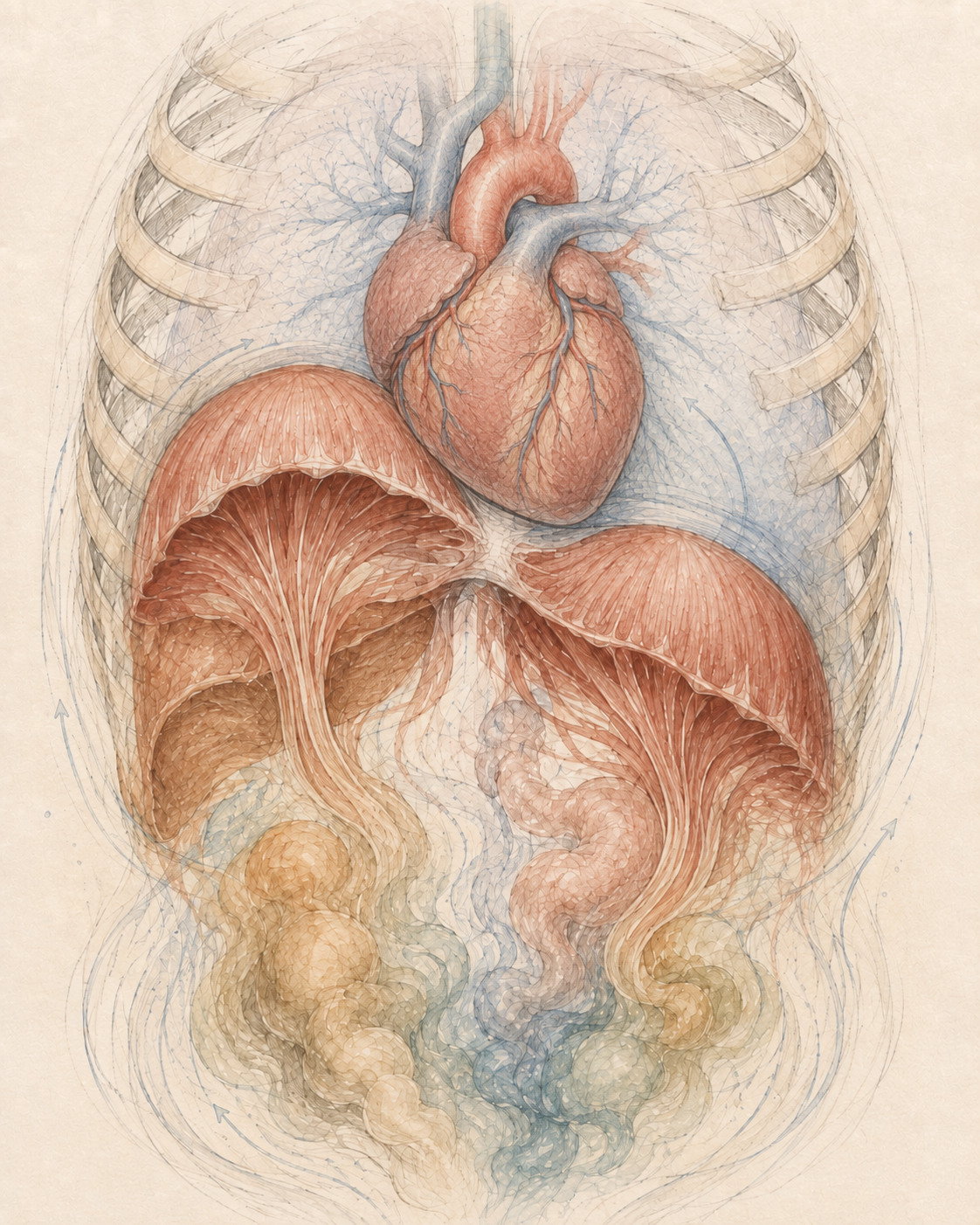
Add in the heart
To give you a visual of the heart in relationship to the moving domes of the diaphragm (represented as two jelly fish)
There aren’t really any neat edges
This is where anatomy always gets much more interesting for me.
In a textbook, everything has a lovely outline.
Here is the diaphragm.
Here is the heart.
Here is the liver.
Here is the kidney.
Here is the muscle.
Here is the fascia.
As though someone has carefully packaged all the parts separately.
But once you start looking at dissection and connective tissue, the idea of those completely separate pieces becomes much harder to maintain.
This is one of the things I enjoy about Gil Hedley’s work.
His dissections encourage us to look not just at individual structures, but at the tissues connecting, wrapping, suspending and separating them. In his work we can see that the body is layered, continuous, slippery, strong, adaptable and that all the structures have relationships.
They live inside membranes and connective tissues.
They slide relative to one another.
They transmit forces.
They share space.
They influence the environment around them.
That does not mean every fascial connection automatically has some enormous therapeutic meaning.
But it does mean anatomy is considerably more interconnected than the diagrams sometimes suggest.
The diaphragm is part of that connective tissue conversation
The diaphragm does not end neatly at the edge of its muscular fibres.
Its surfaces are continuous with layers of connective tissue associated with the thorax, abdominal wall and retroperitoneal region (the deep space at the back of the abdomen, where structures such as the kidneys and adrenal glands sit.)
And that becomes particularly interesting when we start following the diaphragm posteriorly towards the spine.
This is where Thomas Myers’ Anatomy Trains model becomes a useful way of thinking.
In the Deep Front Line model, the diaphragm is considered part of a continuous myofascial pathway that includes structures such as the psoas and pelvic floor.
I want to be very clear here:
Anatomy Trains is a model.
It is not the same thing as saying that the body literally contains a single anatomical cable running uninterrupted from one end of the line to the other.
But models can be useful when they help us ask deeper questions.
And when we get to the diaphragm’s crura, lumbar spine, psoas and quadratus lumborum later in this series, those relationships are going to become very interesting indeed.
That rabbit hole is deepening.
What actually happens when the diaphragm contracts?
At rest, the diaphragm sits in its domed position.
During inspiration, the muscular fibres contract and the shape of the diaphragm changes.
In general, this increases the vertical dimensions of the thoracic cavity and contributes to the pressure changes that draw air into the lungs.
During relaxed expiration, the diaphragm relaxes and returns towards its resting shape as the lungs and chest wall recoil.
That's the simple version which is useful, but it is not the whole story.
Different parts of the diaphragm can move differently.
Its movement changes with: posture, lung volume, rib position, abdominal pressure, muscle activity and of course the task you are doing.
Breathing while lying quietly is not mechanically identical to breathing while walking uphill, lifting a weight, laughing, coughing or running.
So when someone says:
“The diaphragm should move down when I breathe in.”
I think:
Yes.
And what else is happening?
What are the ribs doing?
What is happening posteriorly?
Is the rib cage widening?
What happens at the sternum?
Does one area appear to be doing most of the work?
What happens if we change position?
What does the abdominal wall do?
What happens when the person stops trying to breathe correctly?
That is where breathing assessment becomes much more useful.
It is not just an up-and-down movement
This matters enormously in the way I teach breathing.
If the only instruction we give is:
“Push your tummy out.” or “widen your ribs”
we have reduced a complex three-dimensional event to one visible movement at the front of the body or upper quadrant.
The diaphragm is acting within a rib cage capable of moving in multiple directions.
The lower ribs can widen laterally.
The posterior rib cage can move.
The sternum can respond.
The abdominal contents have to accommodate changes in pressure and position.
So I want breathing to feel much more like space being created in multiple directions than simply the abdomen being pushed forwards or the ribs widened laterally.
This is another place where embodied imagery can be incredibly useful.
Instead of forcing movement, we can begin by imagining it.
The domes changing shape.
The lower ribs widening.
Space opening behind you.
The whole circumference of the lower rib cage participating.
Then we see what the body actually does.
And beneath the diaphragm?
This is where I am going to deliberately leave you hanging.
Because immediately underneath these domes are some extraordinary structures.
The liver.
The stomach.
The kidneys.
And sitting above the kidneys:
the adrenal glands.
They occupy an anatomical and fascial neighbourhood very close to the posterior diaphragm.
There is a genuine relationship there.
But I do not want to turn that into one of those:
“Breathe this way and massage your adrenals and reset your cortisol”
stories.
That is exactly the sort of leap I want this series to avoid.
Later, when we get to the organs, we will look properly at what is connected to what.
What actually moves with respiration.
What fascia is involved.
What the evidence supports.
And where people have taken a real anatomical relationship and run maybe a little too far with it.
Because:
There is a connection.
That does not automatically tell us what the connection does.
The same applies to the heart
You will sometimes hear people say that diaphragmatic breathing “massages the heart”.
It is an appealing image and one I use in teaching to keep it simple, because there is an element of that.
But I think we can do better than that.
There is a real anatomical connection between the diaphragm and the fibrous pericardium.
The position of the heart does change with respiration.
Pressure within the thorax changes during breathing.
Those facts are fascinating.
We do not necessarily need to turn them into a bigger therapeutic claim, although it is tempting.
Amazing anatomy is amazing enough.
And good anatomy should make us both more curious and more precise. Which we will see…
So why does any of this matter when I am teaching?
Because once you begin seeing the diaphragm this way, breathing becomes much harder to teach as an isolated exercise.
When someone breathes in front of me, I am not just watching whether their abdomen rises.
I am looking at the conversation.
What happened to the lower ribs?
What happened behind them?
Did the sternum move?
Did the shoulders have to help?
What happened to their spinal position?
Did they brace?
Did they soften?
Was there movement, or were they trying very hard to manufacture movement?
What happened when they changed their position?
And eventually:
what happened to the abdominal wall and pelvic floor?
Because the diaphragm is not performing its role in isolation.
It cannot.
Its anatomy makes that impossible.
An extraordinary piece of engineering
I called this chapter An Extraordinary Piece of Engineering deliberately.
Although I don't really think of the body as a machine.
The brilliance of the diaphragm is not that it behaves like a clever mechanical pump.
It is that it can change shape and respond while being part of a much bigger living system.
It is muscle and tendon.
It forms two domes.
It moves within the rib cage.
It separates and, simultaneously connects, the thoracic and abdominal spaces.
Its central tendon is firmly connected with the fibrous pericardium surrounding the heart.
It is continuous with wider connective tissue systems.
Important blood vessels and structures pass through it.
It responds to posture, pressure and movement.
And it is doing all of this while you: walk, talk, laugh, cough, lift, run, wee, poo, sing, sleep and live your very individualised gorgeous life.
Most of the time without you consciously thinking about it at all.
That is far more interesting than:
“Take a deep breath into”
And we still haven't reached the edges of the diaphragm.
Explore It in Your Own Body
Reading anatomy is one thing. Experiencing it in your own body is another.
Here are two simple ways to explore some of the ideas from this chapter.
1. Visual Exploration: The Two Jellyfish
Find a comfortable position either sitting, standing or lying down and allow your breathing to settle. There is no need to make your breath bigger or deeper.
Bring your attention to the lower part of your rib cage.
Now imagine two slightly different jellyfish sitting side by side inside your lower ribs.
They aren't identical. The right and left sides of your body have different organs, different shapes and slightly different landscapes around them.
As you inhale, imagine the muscular bells of your two jellyfish contracting and changing shape.
Rather than thinking simply down, picture them becoming a little lower and broader, while the ribs around them make space.
Imagine movement:
to the sides;
towards your back;
and through the whole circumference of your lower rib cage.
Above them, the lungs expand.
Below them, the abdominal contents accommodate that change in shape and pressure.
Then breathe out.
Imagine the muscular fibres softening as the two jellyfish return towards their more rounded, domed resting shape.
Don't try to physically reproduce the picture.
Let the image suggest movement and notice what your body does with it.
Try five or six easy breaths.
Then let the image go.
What changed?
Did you feel movement somewhere you hadn't noticed before?
Did one side feel different from the other?
Did thinking about shape changing feel different from thinking about pushing your abdomen out?
There is no correct sensation to find. The exercise is about curiosity, not performance.
2. Anatomical Exploration: Find the Lower Rib Cage
This time, use your hands.
Place them around the lower part of your rib cage.
Let your thumbs reach towards your back if comfortable, with your fingers resting around the sides and front of the lower ribs.
Don't press hard.
You're simply giving your nervous system another source of information about where your rib cage is.
Take a normal breath in and notice what reaches your hands.
Do the ribs move sideways?
Can you feel anything underneath your thumbs towards the back?
Does the front move more than the sides?
Does one side feel different from the other?
Now breathe out and notice the ribs returning.
Try several breaths without attempting to make them bigger.
Then move your hands slightly maybe further around the sides or further towards the back and explore again.
Remember that you cannot directly feel the diaphragm contracting through your hands.
What you are feeling is the movement of the rib cage and surrounding tissues that accompanies the breathing process.
The aim isn't to force a perfect “360-degree breath”.
It is simply to discover how much of your own rib cage you can notice moving.
Anatomy doesn't just have to be something you know.
It can become something you explore.
Next week: Woven Into the Body
In Chapter Three, we are moving outwards.
We're going to follow the diaphragm into its relationships with the ribs, sternum and lower costal border.
We will look at what actually happens to the rib cage when we breathe.
Why the lower ribs matter so much.
Why breathing should not just be happening at the front of the body.
And why understanding the diaphragm means understanding three-dimensional rib movement.
Because the diaphragm does not move inside a static cage.
The cage moves too.
Welcome further down this particular rabbit hole.
Sources
Standring, S. (ed.). Gray’s Anatomy: The Anatomical Basis of Clinical Practice.
A core anatomical reference for the diaphragm’s structure, domes, central tendon, attachments and relationships with surrounding thoracic and abdominal structures.
Kocjan, J. et al. “Network of breathing. Multifunctional role of the diaphragm: a review.”
A useful review exploring the diaphragm beyond respiration, including its roles in posture, pressure regulation and wider body function.
Further Reading
Franklin, E. Dynamic Alignment Through Imagery.
A useful introduction to using imagery to understand and experience anatomy as something dynamic and moving, rather than fixed and static.
Myers, T. Anatomy Trains: Myofascial Meridians for Manual Therapists and Movement Professionals.
Useful for exploring the diaphragm within a wider myofascial model, particularly its relationship with the psoas, deep front line and pelvic floor. Best understood as a model for thinking about continuity rather than as a literal anatomical map.